胸部X-ray判讀基礎
出自KMU Wiki
| 在2007年11月30日 (五) 15:38所做的修訂版本 (編輯) Terrasa (對話 | 貢獻) ←上一個 |
在2007年11月30日 (五) 15:47所做的修訂版本 (編輯) (撤銷) Terrasa (對話 | 貢獻) 下一個→ |
||
| 第1行: | 第1行: | ||
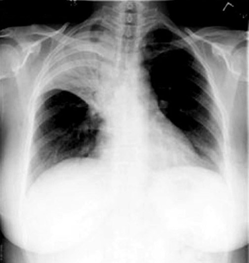
| - | <blockquote dir="ltr">[[Image:pleural effusion]]胸部X-ray判讀基礎~~</blockquote> | + | <blockquote dir="ltr">[[Image:Pleural effusion|Image:pleural effusion]]胸部X-ray判讀基礎~~</blockquote> |
| 一.判讀X光片:'' 首先要查看片子的基本資料:姓名.照射日期.姿勢(A-P View,''' P-A View or Lateral View)品質 (KV+Density). | 一.判讀X光片:'' 首先要查看片子的基本資料:姓名.照射日期.姿勢(A-P View,''' P-A View or Lateral View)品質 (KV+Density). | ||
| 第40行: | 第40行: | ||
| | | | | ||
| |- | |- | ||
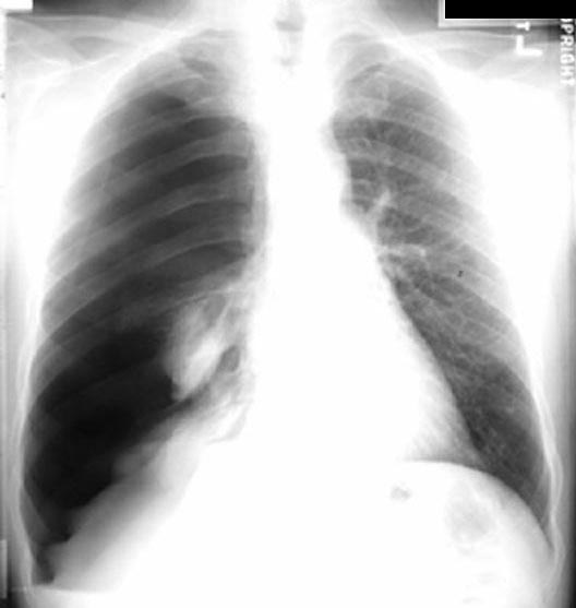
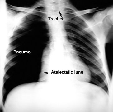
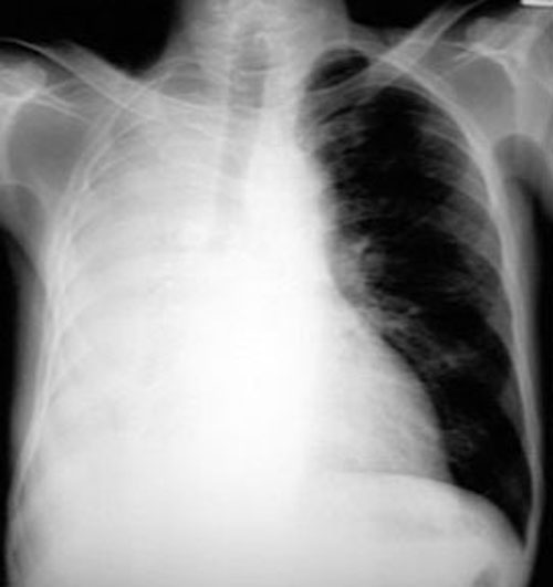
| - | | | + | | [[Image:Relaxation_atelectasis.jpg]] |
| - | | | + | | [[Image:Lung_collapse.jpg]] |
| |- | |- | ||
| | | | | ||
在2007年11月30日 (五) 15:47所做的修訂版本
Image:Pleural effusion胸部X-ray判讀基礎~~
一.判讀X光片: 首先要查看片子的基本資料:姓名.照射日期.姿勢(A-P View,' P-A View or Lateral View)品質 (KV+Density).
二.評估胸部X光成像質:
**技術品質Technical quality
1.Density orX-ray penetration :透過Heart陰影必須可見到Spinal cord.
2.Position of the Scapulae:肩胛骨的位置應置於肺野之外.
3.Rotation of the CXR(採正面):兩側鎖骨近胸骨內側端應與Spinous process等距.
4. Degree of inspiration(吸氣末憋氣):Anterior segment of the 6th rib
Posterior segment of the 9th rib
三.如何判讀X-ray:
1.Airway
2.TheHila:Hilum左邊的肺門高於右側 ;肺門的位置可因Lung collapse, Lung fibrosis or Pneumonia change.
'3.Normal Lung Markings:Lung markings is Lung Vessels on X-ray present.
4.Diaphragm:右邊高於左邊,因右邊有Liver關係, 右側Diaphragm最高點在內1/2處.
5.Costophenic angle(C-P angle).
6.Heart:RA,SVC,Aortic Arch,LA,LV and Pulmonary Artery;Normal Heart size<50%;Cardiomegaly>50%.
7.Skull.

| 
|

| |

| 
|
